Equine Cervical Spinal Surgery
Why do Spinal Surgery?
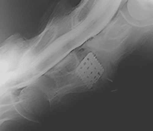
The myelogram has shown that your horse has spinal cord compression at one or more levels. Why do cervical spinal surgery? The surgical technique currently used in equine cervical spinal surgery is designed to fuse the articulation (intervertebral space), thereby stopping the movement of the affected cervical spinal joints that compress the spinal cord when the neck is flexed or extended. The fusion process also allows the spinal canal to enlarge by having the enlarged articular facets to atrophy (shrink in size) as with the fusion, and they are no longer needed.
Without Spinal surgery, excessive movement of the cervical vertebra continues to damage the spinal cord, and after a period of time, depending on the severity of the spinal cord compression, the damage to the spinal cord will become irreversible and not responsive to surgery.
It is not known how long this will take, but we do know that horses operated on within 30 days of the onset of clinical signs will have a more favorable outcome.
Surgeons are trying to encourage early surgical operation by discounting surgical fees for horses operated on within 30 days of the myelogram.
Cervical Spinal Surgery
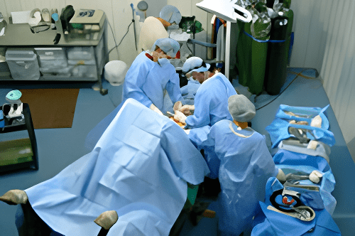
Cervical Spinal surgery is done with the horse on his back, dorsal recumbency, under general anesthesia. The front legs are tied back to help get the shoulder muscles out of the way and expose the cervical spine, especially for C6/7 and C7/T1.
A special neck brace will hold the neck in position, and an image cassette will be in place during surgery. The neck, head, and body positioning are critical for proper implant placement.
Once the horse is on the table in the proper position, he/she is prepped and draped for surgery. An incision is made, and the spine is exposed. A small portion of bone is chiseled off.
Marking pins are gentled placed, and an intraop image is taken to help locate the vertebra. The marking pins will assist in giving the proper angle for the drill guide and, ultimately, the implant. A series of instruments, drills, core saws, and kerf cutters are used to prepare the hole for the implant.
Throughout the surgery, intraoperative images are taken to help evaluate the depth of the hole for the implant. Once the hole is finished, it is tapped and cleaned for the placement of the implant. The implant is twisted into place, again with the help of intraoperative imaging, to make sure the implant is at the proper depth.
Bone marrow, harvested from the hole, is used to fill the implant and speed up the fusion process. After the implant is filled with bone marrow, the muscles are sewn back together. Staples are used on the skin to close the incision and followed with a short-term recovery bandage.
The surgery time for placing one implant, a single level, can be from 45 minutes to 1 hour. Whereas placing two implants, a double level, can be done in 1.5 to 2 hrs. The location of the lesion, the age of the horse, and the intraop radiograph equipment are just a few factors that can change the operating time.
The horse is taken to recovery, where it can last 1-2 hours, depending on factors such as length of anesthesia and grade of wobbler. It is always best for the horse to take at least 45 minutes to recover. This is usually a very long 45 minutes – 1 hour for the owner as compared to the surgery time.
When the horse is up, a new compression bandage is placed before moving to his/her stall. The bandage will stay on for 5-7 days and can be changed as needed.
To watch a spinal fusion surgery, click on this link: Spinal Fusion Presentation.
Spinal Surgery
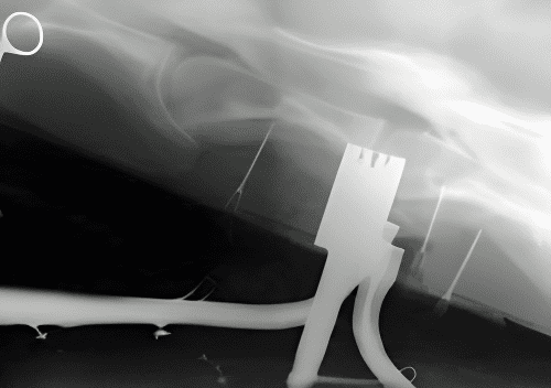
Intra-op image showing kerf cutter
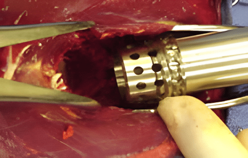
Placing the Seattle Slew Implant
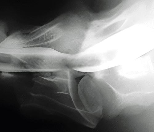
Myelogram showing compression