Myelogram/CT
Why do a Myelogram?
A myelogram is a diagnostic tool used to determine cord compression in the potential surgical candidate. Even when the horse shows obvious compression on standing films, the myelogram is needed to show if there is more than one level of compression. Cord compression is determined by measuring the dorsal and ventral dye columns in both neutral and flexed positions. Ratio measurements are made by using a special software program on each digital radiograph taken during the myelogram.
What is a myelogram, and how is it performed?
A myelogram is performed under IV anesthesia to check compression of the spinal cord. With the widespread use of digital radiography, it is not always necessary to refer to a university or referral center. A myelogram involves an injection of an iodine-based fluid into the spinal canal that will outline the spinal cord, replacing the clear radiolucent cerebrospinal fluid when viewed by radiographs.
The myelogram is accomplished by carefully inserting a spinal needle into the space between the first and second cervical vertebra (A-O space). Feeling a "pop" indicates penetration of the protective membrane (dura mater). The CSF (cerebrospinal fluid) is withdrawn through a long extension tube, and then the dye is injected over a three-minute period of time. A series of radiographs are then taken with the cervical vertebra in neutral, flexed, and extended positions.
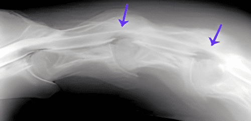
Compression needs to be at least a 50% reduction in width when compared to the width of the dye column in a neutral position.
After reviewing the myelogram, your horse might show a single spinal canal compression. This is referred to as a "single level" and will require one Seattle Slew implant. Two compressions would be considered a "double level," requiring two Seattle Slew implants. If your horse has a negative myelogram and no compressions, it is still worth the time and effort for the client and patient.
To get a better visual understanding of how a myelogram is done, you can watch a myelogram presentation on SeattleSlew.com.
Click here for information on a nuclear scan.
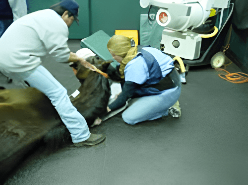
Myelogram Procedure
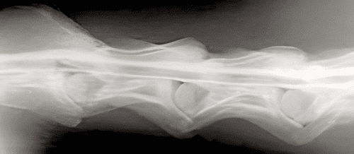
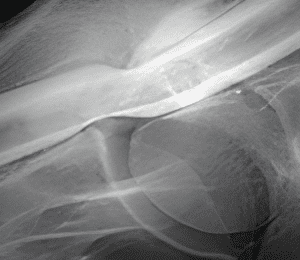
Neutral view with dye around
Flexed view with dye around
Myelogram showing compression1
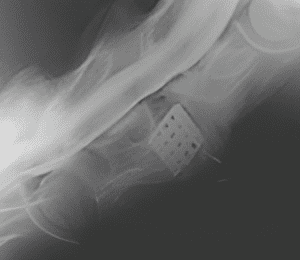
Myelogram showing implant with no compression
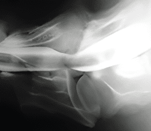
Myelogram showing enlarged picture of a compression
Computed Tomography aka “CAT SCAN”
In the last 10 years newer technology has become available that will allow a Computed tomography procedure to be performed on the caudal cervical and anterior thoracic area of very large horses. This mode of imaging has been available for humans and smaller animals for over 25 years but the equipment has not been large enough to allow visualization of the caudal neck of a large horse. While a myelogram can detect compression of the spinal cord in a dorsal/ventral plane it is not able to show compression from side to side. A CT scan can do that and allows us to see areas of spinal cord compression/arthritis/rib fractures and soft tissue masses such as melanomas. See the video attached.
A myelogram combined with a CT scan (performed at the same time) is now the gold standard for equine centers working on the diagnosis and treatment of caudal cervical cord compression. If you are interested in knowing more contact a surgeon located in the contact list.


