Standing Cervical Radiographs
Cervical Radiographs Following a Neuro Exam
After a neurological examination is completed, the next step will be standing cervical radiographs to help evaluate the cervical spine for fractures, congenital malformations, arthrosis, subluxation, and a narrow spinal canal.
The standing cervical radiographs can be done in the field with a portable digital x-ray unit. With the new digital units, the images are of excellent quality. Your horse can also be taken to a clinic/hospital environment with a large x-ray unit. The radiographs usually require a small amount of sedation to get the horse to stand still for the series of exposures.
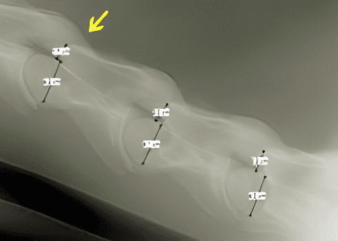
The series of radiographs starts at the base of the head, as shown in the "Cassette Placement" picture to the right.
Picture "A" will show the AO (atlas occipital joint) to C2/C3.
Picture "B" will show C2 to C4.
Picture "C" will show C4 to C7. Usually, this view is taken from both the left and right sides of the horse.
C7 is more easily seen with the leg closest to the generator elevated and pulled posteriorly.
Depending on the size of the horse, a complete series will include 4–5 images to cover the whole vertebra.
After the radiographs are taken, they can be examined for fractures, congenital malformations, arthritis of the articular facet joints, subluxation, narrowing of the spinal canal, and degeneration of the disc space. These conditions do not always result in compression of the spinal cord, but can be an indication of possible spinal cord compression.
Using a software program with the radiographs, the cord ratio measurements are taken. These ratios help in the objective assessment of the narrow spinal canal.
If a compression is suspect from these ratios, then a myelogram must be performed for further diagnostics.
Click here to learn more about the myelogram process.
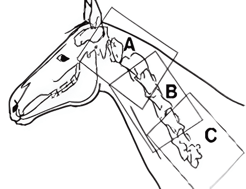
Cassette Placement